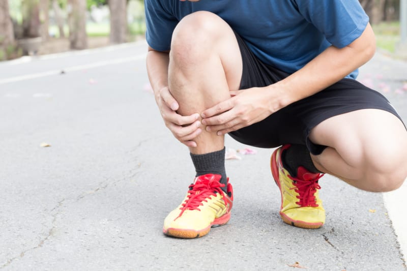
With marathon season fast approaching many are in training mode, and one of the most common injuries I see at this time in my clinic is medial tibial stress syndrome (MTSS). This commonly gets called ‘shin splints’ which is a catch-all term to explain leg pain on exercise and is not a diagnosis. There are many diagnoses for exercise-induced leg pain, so it’s important that you obtain the correct analysis to get the right treatment and get you back to sport quickly.
This blog aims to give a brief update of what MTSS is, how to spot it and what to do about it, with some self-treatment advice at the end.
So what is MTSS?
Well, the truth be told we are still not entirely sure, and the most current definition we have is ‘Pain felt along the middle or distal third of the posteromedial border of the tibia that occurs during exercise, excluding pain from ischemic origin or signs of stress fracture (Yates & White, 2004). Currently, there are 2 schools of thought: the anatomical and the bone stress biomechanical theories.
Anatomical Theory:
The anatomical theory proposes that certain muscles within the leg contract and pull against the tibia and the surrounding tissue called the periosteum, causing symptoms; although, we are not 100% sure which muscle is to blame. There is research supporting all of the muscles in the deep compartment of the leg being to blame, most probably because we are all different (or technically, have anatomical variation). Whilst many of us conform to ‘normal’ anatomy, variation does exist, and this can make the results of the research appear confusing (Stickley, Hetzler, Kimura, & Lozanoff, 2009).
Bone Stress / Biomechanical Theory:
The next school of thought is the biomechanical principle and which really looks at the amount of stress going through the bone and the bones reaction to this stress. As you apply a force perpendicular to the leg bone (tibia), it will bend at the narrowest point (this is termed a bending moment), and this point tends to be the site at which MTSS symptoms occur, as shown in the picture below.
As bone is living tissue, it responds to stress, and we know that bone needs to be stressed to remodel and grow new bone. Injuries occur when too much stress is applied over a prolonged period of time, and eventually, the bone reaches failure point, which can result in a stress fracture.
There was some thought that if MTSS were left untreated, it would eventually lead to a stress fracture putting MTSS and stress fractures on the same continuum. However, this may not be the case as not everyone with MTSS goes on to develop a stress fracture. Furthermore, there is no clear evidence that people with stress fractures had MTSS prior to the stress fracture.
A mix of both theories:
There is also some thought that actually MTSS is a combination of the anatomical and the bone stress theories. One study demonstrated that as a muscles fatigues, the bone stress increases, as the muscles are unable to oppose the bending moments in the tibia (Milgrom et al., 2007). In my opinion, this would appear to be a reasonable explanation.
How does MTSS present?
The good thing about MTSS is that it has a nice clear presentation which includes:
- Pain on running (can occur on fast pace walking) which initially does not cause you to stop running; however, this may be the case if symptoms and activity continue.
- The pain is normally described as an intense ache.
- On palpation there is pain along the lower inside border of the shinbone (tibia), this is known as the lower medial third of the tibia.
- After running the pain settles within 48 hours and does not wake you up at night.
What causes MTSS?
There are many risk factors for MTSS, with no one factor regularly to blame. MTSS is known as a multifactorial pathology which means that multiple factors are contributing to the problem. From the research, we can see some of the most common causes are:
- Too Much Too Soon. Bone takes time to adapt to new stress levels. MTSS is very common in people who have come back from injury or previously did little activity, and try to do too much too soon.
- The Female Sex. Unfortunately, women are at higher risk of developing bone stress injuries compared to men due to what is known as the female triad, which reduces bone strength. There are 3 elements to the triad: irregular periods, osteoporosis (reduced bone density) and low-energy consumption (poor, low-calorie diet). Clearly, this does not apply to all females, and it is important to remember that men can get stress-related injuries as well.
- Excessive Foot Pronation is when the foot rolls in (pronates) too much and too quickly, resulting in increased stress going through the tibia and increasing the bending moment, hence increased stress. It may also result in increased muscle activity and thus stress to the bone.
- Poor Proximal Control is an area that is frequently overlooked and forgotten about when, actually, it is very important. Evaluation of the function at the hips and pelvis when running can reveal areas of poor function: this may be reduced motion, weakness or stiffness. One common weakness is within the gluteal muscles, which results in internal hip/knee rotation and foot pronation. This is where detailed gait analysis comes into its own. The secret to successful management is a clear identification of the cause of the problem. By analysing the pelvis, hip, knee and foot, we can determine where the site of dysfunction is occurring and develop an individual management plan (see below under gait analysis).
- Calf muscle weakness. We know that in people with MTSS, the functional calf strength is weaker than those without. Whilst focusing on Gastrocnemius strengthening is good, the Soleus muscle should be targeted as well.
- Tibial Varum is the natural bowing in the leg which is compensated by foot pronation and tends to increase the amount of bending that occurs during running.
How to treat MTSS
No one treatment works for everyone. Due to the multifactorial nature of MTSS, it is common that a range of interventions is required. A successful treatment plan can only be provided once the cause of the pain has been determined. For example, if MTSS is being caused by excessive foot pronation and calf and gluteal weakness, all of these must be addressed to treat and prevent further pain.
Common treatments include:
- Foot orthoses (insoles) are used to try to reduce bone stress. There is good evidence to support the use of orthoses, although the research is confusing as to which type of orthoses are most appropriate (Craig, 2009; Loudon & Dolphino, 2010; Moen, et al., 2009; Reshef & Guelich, 2012, Rome, et al., 2005; Yeung, Yeung, & Gillespie, 2011). The most likely explanation for this is the variability between individuals and the need for tailored prescriptions-just as eyeglasses will only be effective with the right prescription, foot orthoses are the same.
- Running technique. There is evidence that reducing stride length, increasing the base of support (width between the feet), and increasing cadence can all reduce the stress through the tibia. Simple running cues can help to change your running technique. Gait analysis can be helpful to offer some visual guidance and also to show a before and after effect (Franklyn-Miller, A., Roberts, A., Hulse, D., & Foster, J. 2012).
- Gait Analysis is very useful in picking up on poor function and guiding an effective treatment plan. Gait analysis ranges from a simple visual analysis, a 2-D method (using video cameras), in-shoe pressure measurements (using force sensing insoles inside the shoe) or 3-D analysis. To obtain kinematic data (a measure of movement), particularly joint rotation, 3-D analysis is required; though, just altering the kinematics may not reduce the pain.
- Strength and Conditioning. A very interesting article was published in the British Journal of Sports Medicine, showing that strength and conditioning help prevent injuries by 68% (Lauersen, Bertelsen and Anderson, 2013).
- Proprioception is our sense of balance and joint position. After an injury, there is weakness surrounding the area, as well as the reduced ability of the foot’s stretch receptors to send messages to the brain. As a result, there is less appreciation of the position of the foot and an increased risk of further injury. Proprioception is often forgotten about in assessments and treatment of injuries; however, an article in the British Journal of Sports Medicine showed that proprioception training reduced injuries by 45% (Lauersen, Bertelsen and Anderson, 2013).
- Footwear advice may be required to help with MTSS. Depending on the individual, they may require increased cushioning or increased stiffness (for stability). The evidence surrounding running shoes is very weak, and the traditional methods of advising on the type of shoe have no scientific support. By measuring the degree of pronation on 3D gait analysis, we can have a more objective indication of the most appropriate shoe for an individual. One clear thing is that comfort is very important. The most comfortable shoe for the individual is generally the one in which there is the least risk of injury.
What can I do at home?
Calf strengthening is very simple and can be done at home.
Gastrocnemius strengthening involves going up on tiptoes and back down to heel contact. Once you are able to do 30 of these without feeling any pain or tightness in the calf muscle, progress to one leg only. You can progress this further by increasing the resistance.
For Soleus strengthening, the heel raises are completed with the knees bent, and the same protocol is followed.
Summary
As you will now appreciate, Medial Tibial Stress Syndrome is a very complex, multi-factorial pathology. It is key to find the right treatment program for you, as one treatment on its own is not often enough to settle the symptoms. Treatments are tailored to you, and with the right treatment, it is a condition that can be prevented, allowing you to get that personal best at this year’s London Marathon.
Best of luck,
Nick
Nick Knight is a Sports Podiatrist. You can find him at our Moorgate clinic. To make an appointment call 020 7482 3875 or email info@complete-physio.co.uk.
References
Craig, D. I. (2009). Current developments concerning medial tibial stress syndrome. Phys Sportsmed, 37(4), 39-44. doi: 10.3810/psm.2009.12.1740
Franklyn-Miller, A., Roberts, A., Hulse, D., & Foster, J. (2012). Biomechanical overload syndrome: defining a new diagnosis. British journal of sports medicine, bjsports-2012.
Lauersen, J. B., Bertelsen, D. M., & Andersen, L. B. (2013). The effectiveness of exercise interventions to prevent sports injuries: a systematic review and meta-analysis of randomised controlled trials. British journal of sports medicine, bjsports-2013.
Loudon, J. K., & Dolphino, M. R. (2010). Use of foot orthoses and calf stretching for individuals with medial tibial stress syndrome. Foot Ankle Spec, 3(1), 15-20. doi: 10.1177/1938640009355659
Milgrom, C., Radeva-Petrova, D. R., Finestone, A., Nyska, M., Mendelson, S., Benjuya, N. Burr, D. (2007). The effect of muscle fatigue on in vivo tibial strains. J Biomech, 40(4), 845-850. doi: 10.1016/j.jbiomech.2006.03.006
Moen, M. H., Tol, J. L., Weir, A., Steunebrink, M., & De Winter, T. C. (2009). Medial tibial stress syndrome: a critical review. Sports Med, 39(7), 523-546. doi: 10.2165/00007256-200939070-00002
Reshef, N., & Guelich, D. R. (2012). Medial tibial stress syndrome. Clin Sports Med, 31(2), 273-290. doi: 10.1016/j.csm.2011.09.008
Rome, K., Handoll, H. H., & Ashford, R. (2005). Interventions for preventing and treating stress fractures and stress reactions of bone of the lower limbs in young adults. Cochrane Database Syst Rev(2), Cd000450. doi: 10.1002/14651858.CD000450.pub2
Stickley, C. D., Hetzler, R. K., Kimura, I. F., & Lozanoff, S. (2009). Crural fascia and muscle origins related to medial tibial stress syndrome symptom location. Med Sci Sports Exerc, 41(11), 1991-1996.
Yates, B., & White, S. (2004). The incidence and risk factors in the development of medial tibial stress syndrome among naval recruits. Am J Sports Med, 32(3), 772-780
Yeung, S. S., Yeung, E. W., & Gillespie, L. D. (2011). Interventions for preventing lower limb soft-tissue running injuries. Cochrane Database Syst Rev(7), Cd001256. doi: 10.1002/14651858.CD001256.pub2
Don’t let pain hold you back, book now!


